Product Overview
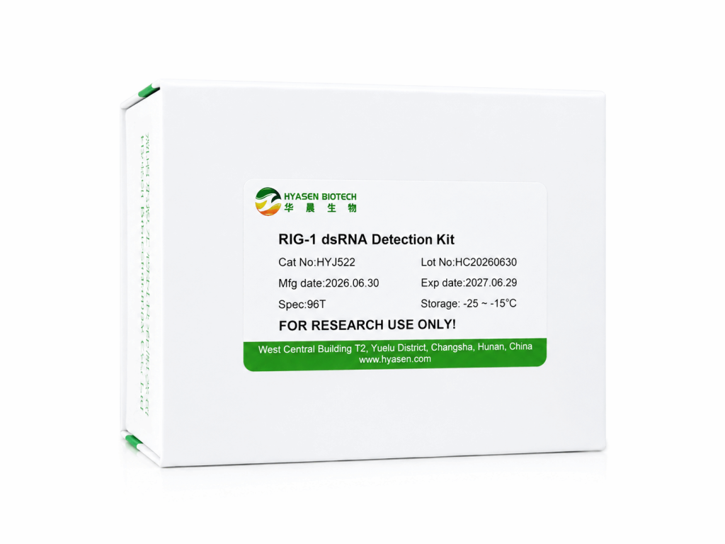
RIG-I (retinoic acid-inducible gene I) is an RNA-sensing receptor whose ATPase activity is stimulated by interactions with double-stranded RNA. The Hyasen RIG-I dsRNA Detection Kit uses this biochemical response to investigate dsRNA in RNA samples. During the first stage, RIG-I is incubated with ATP and the sample; dsRNA-dependent activity produces ADP. A second reagent depletes remaining ATP, and a final conversion/detection reaction converts ADP back to ATP for luciferase/luciferin-based luminescence measurement. A standard curve is used to estimate dsRNA content. Unlike an ELISA, this assay measures receptor-associated enzymatic activity rather than antibody capture. It can therefore provide a complementary readout when characterizing RNA impurities in mRNA research workflows.
Components
| Components | 96 T | 384 T | |
| RIG-I ATPase Reaction | RIG-I enzyme(1µM) | 100µL | 400µL |
| 10× RIG-I Reaction Buffer | 0.25mL | 1mL | |
| 1M DTT | 20μL | 50μL | |
| 10mM ATP | 25μL | 100μL | |
| Standard (UTP, 25ng/μL) | 40μL | 150μL | |
| Standard (N1-Me-pUTP,25ng/μL) | 40μL | 150μL | |
| Measurement of ADP | Reagent 1-Assay buffer | 0.5mL | 2mL |
| Reagent 1-Enzyme(5×) | 0.125mL | 0.5mL | |
| Reagent 1-0.5M MnCl2 | 25μL | 100μL | |
| Reagent 2 | 1.25mL | 5mL | |
| 384-well Plate | 1 plate | 1 plate |
Product Features
- RIG-I ATPase-based detection principle, distinct from antibody-based ELISA.
- Includes UTP- and N1-Me-pUTP-dsRNA standards for calibration.
- Small-volume workflow: 5 µL of standard or sample is combined with 5 µL of RIG-I/ATP mix.
FAQ
Q1: How does this RIG-I-based assay differ from antibody-based dsRNA detection methods, such as ELISA?
Antibody-based methods, such as ELISA, detect dsRNA based on structural recognition by anti-dsRNA antibodies, independent of biological function. This RIG-I dsRNA Detection Kit instead measures the functional activation of RIG-I’s ATP hydrolysis activity upon dsRNA binding — the same biochemical event that triggers innate immune signaling in cells — providing a readout more directly linked to the immunogenic potential of the dsRNA present in a sample.
Q2: What is the purpose of the ATP depletion step (Reagent 1) in the detection workflow?
After the RIG-I reaction, residual unreacted ATP remains in the sample alongside the ADP generated from dsRNA-triggered hydrolysis. Since the final detection step relies on converting ADP into ATP for luminescent quantification, any leftover unreacted ATP would create background signal and compromise accuracy. Reagent 1 depletes this residual ATP, ensuring that the subsequent luminescent signal specifically reflects the ADP generated by the RIG-I reaction.
Q3: What sample volume and equipment are needed to run this assay?
The assay is compatible with reaction volumes as low as 5 μL, performed in a standard multi-well plate format. Detection requires a luminescence-capable plate reader to measure the luciferase/luciferin signal generated in the final detection step; dsRNA content is then calculated by comparing sample luminescence values to a standard curve generated using known dsRNA concentrations.