Product Overview
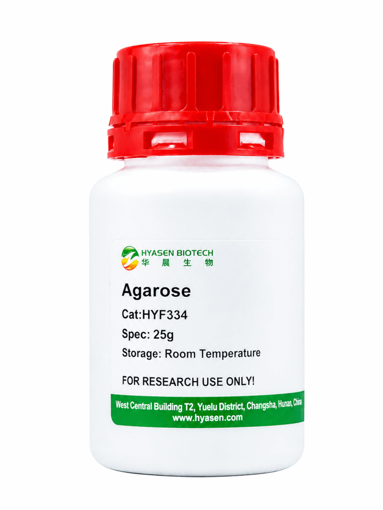
HYF334 Agarose is a high-purity agarose designed for nucleic acid separation by gel electrophoresis. Agarose purity can influence DNA band resolution and the clarity of electrophoresis results, making it an important consideration in routine molecular biology workflows.
Agarose is commonly prepared as a gel at concentrations ranging from 0.5% to 2%, depending on the nucleic acid fragments to be analyzed and the desired separation characteristics. The resulting gel matrix enables the separation and identification of DNA fragments and supports applications such as DNA analysis and restriction enzyme mapping.
For visualization, a suitable nucleic acid fluorescent stain can be incorporated into or applied to the gel according to the staining protocol. Following electrophoresis, DNA bands can be observed using a compatible gel imaging system.
HYF334 Agarose is suitable for routine laboratory workflows that require agarose gel preparation, DNA fragment separation, and nucleic acid analysis.
Product Features
- High-Purity Gel Matrix: Designed for nucleic acid electrophoresis, where agarose purity is an important factor affecting DNA band resolution and result clarity.
- Flexible Gel Concentrations: Suitable for preparing agarose gels at typical concentrations of 0.5–2%, allowing users to adjust gel concentration according to their separation requirements.
- Broad DNA Analysis Applications: Supports DNA fragment separation and identification, restriction enzyme mapping, and routine nucleic acid analysis using compatible fluorescent stains and gel imaging systems.
FAQ
Q1: What is HYF334 Agarose used for?
HYF334 Agarose is used to prepare gels for nucleic acid electrophoresis. It supports DNA fragment separation and analysis, including DNA identification and restriction enzyme mapping.
Q2: What agarose concentration should I use for DNA electrophoresis?
Agarose gels are commonly prepared at concentrations of 0.5–2%. The appropriate concentration depends on the DNA fragment sizes and the desired separation resolution. Select the concentration according to your sample and electrophoresis protocol.
Q3: How can DNA bands be visualized after agarose gel electrophoresis?
Use a suitable nucleic acid fluorescent stain according to its instructions. After electrophoresis, visualize the DNA bands with a compatible gel imaging system. Follow the stain manufacturer’s safety and handling recommendations.