Product Overview
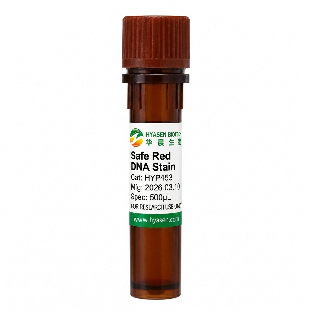
The HYP453 Safe Red DNA Stain is a concentrated fluorescent dye designed for DNA detection in agarose gel electrophoresis. Its 10,000× stock formulation is compatible with commonly used electrophoresis buffers, including TAE, TBE, and rapid electrophoresis buffer (RRB), making it suitable for routine nucleic acid analysis workflows.
With a reported molecular weight of approximately 1,240 g/mol, the dye is designed to have limited cell membrane permeability. It provides sensitive DNA visualization and can serve as an alternative to conventional ethidium bromide (EB) staining under suitable experimental conditions.
Safe Red DNA Stain has spectral characteristics similar to EB, with strong excitation in the 250–300 nm range. UV-based gel imaging systems are therefore recommended for DNA band visualization. The product can be stored at room temperature for up to two years when protected from light.
Product Features
- Sensitive DNA Visualization: Designed for fluorescent detection of DNA in agarose gels, offering an alternative to conventional ethidium bromide staining.
- Concentrated 10,000× Formulation: Compatible with TAE, TBE, and RRB electrophoresis buffers, supporting flexible use across common gel electrophoresis workflows.
- Suitable for UV Gel Imaging: Its spectral characteristics are similar to those of ethidium bromide, with strong excitation in the 250–300 nm range for imaging on compatible UV-based gel documentation systems.
FAQ
1. What is Safe Red DNA Stain used for?
It is used to fluorescently stain and visualize DNA during agarose gel electrophoresis, supporting routine molecular biology applications such as DNA fragment analysis.
2. Which electrophoresis buffers are compatible with Safe Red DNA Stain?
The stain is compatible with TAE, TBE, and rapid electrophoresis buffer (RRB). Follow the recommended staining protocol for your specific workflow.
3. Which imaging system is recommended for Safe Red DNA Stain?
A UV-based gel imaging system is recommended because the stain has strong excitation in the 250–300 nm range and spectral characteristics similar to ethidium bromide. Follow appropriate UV exposure and laboratory safety procedures.