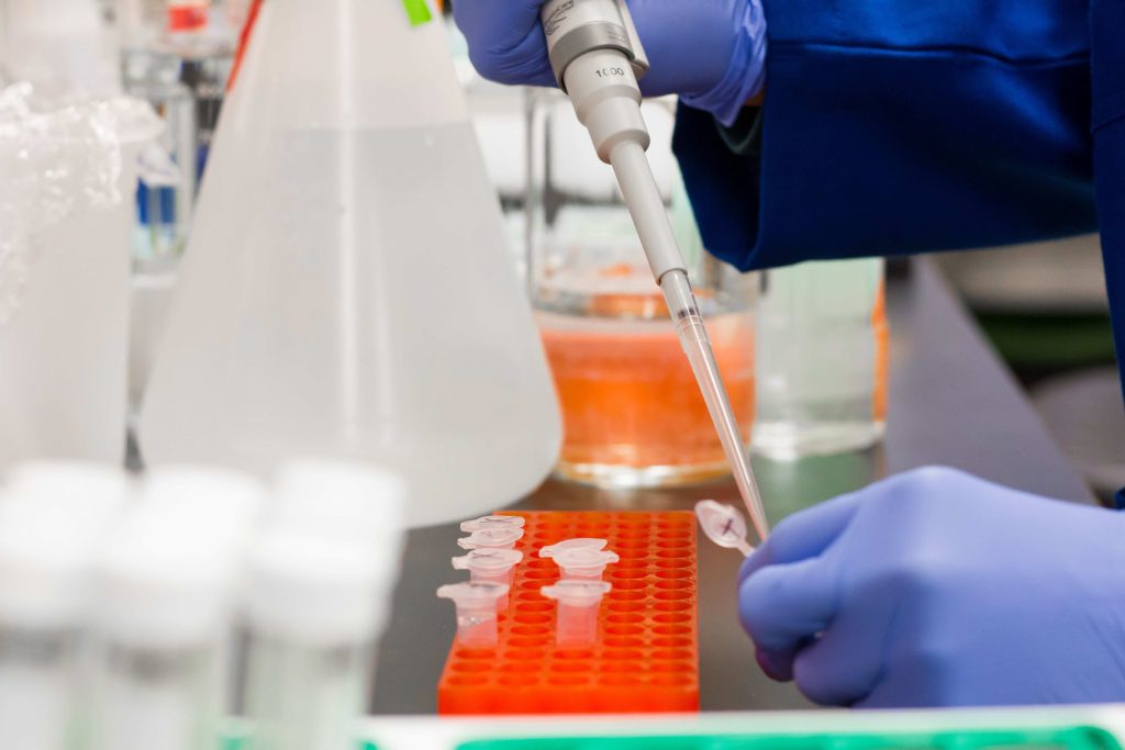
A DNase activity assay measures how quickly a DNase sample degrades a defined DNA substrate, usually by tracking fluorescence in a microplate reader and reporting the result in activity units.
For biopharmaceutical manufacturers that number is operational, not academic. DNase is added on purpose — to digest the DNA template after in vitro transcription, to reduce nucleic acid load during purification — and then becomes a process-related impurity that must be shown to be removed. A DNase activity assay is how that control is demonstrated, and how incoming water, buffers and reagents are screened before they reach a GMP step.
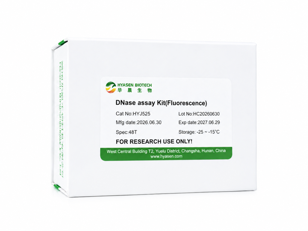
This guide covers the fluorescence format: its principle, how results are read and quantified, how to validate the method under ICH Q2(R2), and what to check when selecting a kit. The format described here is the one used in our DNase Assay Kit (Fluorescence) (HYJ525).
Key takeaways
- A fluorescence DNase activity assay uses a quenched DNA probe: cleavage releases the fluorophore, so rising signal tracks enzyme activity (Ex/Em 485/525 nm).
- Screening reads a ratio — RFU30 ≥ 2 × RFU0 indicates contamination. Quantification uses a standard curve from 1.25×10⁻⁶ to 1×10⁻⁵ U/μL with r ≥ 0.99.
- Unit definitions differ between suppliers. In the HYJ525 manual, 1 unit (1 µg pBR322 DNA fully degraded in 10 min at 37 °C) equals 0.3 Kunitz units.
- For regulated use, treat the kit as a method to be validated: matrix specificity, spike recovery, precision, LOQ and robustness all need documented evidence.
What Is a DNase Activity Assay?
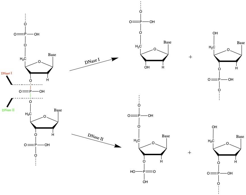
A DNase activity assay is an analytical procedure that quantifies enzymatic degradation of DNA: a defined substrate is incubated with the sample under fixed buffer and temperature conditions, and the rate of substrate loss is converted into activity units.
Two things separate a usable assay from a misleading one. The substrate and conditions must be fixed, because DNase I acts differently on double-stranded DNA, single-stranded DNA and DNA:RNA hybrids, and differently again at acidic versus neutral pH. And the unit definition must be known, because “1 unit” is a manufacturer-specific statement unless it references a published definition.
In practice the assay answers three questions: is this material contaminated; how much active nuclease is present; and is the residual level below the validated limit.
Why Biopharma Manufacturers Test DNase Activity
Nuclease is treated as a process-related impurity rather than a background nuisance. ICH Q6B requires process-related impurities to be identified, quantified and controlled to a justified limit [1]. The Chinese Pharmacopoeia 2020 general chapters for human vaccines, recombinant monoclonal antibodies and gene therapy products each require nuclease residues to be tested in an appropriate intermediate when the finished product cannot be tested directly [2].
For mRNA products, WHO regulatory considerations describe the process explicitly: after transcription the DNA template is digested with DNase before purification, and removal of production enzymes — including DNA polymerase and restriction enzymes — must be demonstrated [3]. Residual DNA is separately capped at 10 ng per parenteral dose for continuous cell lines [4], which is one reason nuclease treatment sits inside a validated, documented step. Reagents for this stage, including the nuclease itself, are listed in our biopharma solutions overview.
| Process point | Why DNase activity is measured |
| Incoming water, buffers and raw materials | A contaminated buffer can degrade intermediates across a batch; screening prevents that |
| IVT template digestion (mRNA) | Confirms the DNase step worked and supports the template-removal claim |
| Viral vector and plasmid purification | Verifies that added endonuclease is removed or inactivated to the validated level |
| Cleaning verification | Confirms surfaces and equipment are free of detectable nuclease after cleaning |
| Enzyme lot release (supplier side) | Assigns reproducible units to each lot so “1 unit” means the same across shipments |
Note the two different questions behind these rows. Contamination screening is binary — is any activity detectable? An intentionally added nuclease is quantitative and two-sided: enough activity to do the job, low enough residual afterwards. One format can serve both, but sample preparation, dilution and acceptance criteria must be defined separately. Labs that also prepare RNA in-house meet the same contamination logic at the bench; our RNA purification protocol guide covers nuclease-free handling for those workflows.
How a Fluorescence DNase Activity Assay Works
Fluorescence formats replace the classical absorbance readout with a quenched oligonucleotide probe. The probe holds a fluorophore and a quencher in close proximity, so the intact probe emits little signal. When DNase cleaves the probe, fluorophore and quencher separate and fluorescence increases [5].
The signal therefore moves opposite to the substrate: the more activity, the faster fluorescence rises. In the HYJ525 format the reaction is read at Ex/Em = 485/525 nm, and the rate of increase is positively correlated with the amount and activity of the enzyme [6].
Three practical consequences follow. A 30-minute incubation at 37 °C is enough for a qualitative call, so the assay fits in-process testing. Background is low because the substrate is a dedicated probe rather than bulk genomic DNA, which pushes detection into the 10⁻⁶ U/μL range. And the same wells can be read as an endpoint (RFU0 and RFU30) or kinetically at 1–1.5 minute intervals when the reader supports kinetic mode [6].
DNase Activity Assay Methods Compared
| Method | Readout | Quantification | Strength | Limitation |
| Kunitz UV absorbance (A260) | Absorbance increase from hydrolysis | Quantitative, Kunitz units | Reference method, no special reagents | pH 5.0, high enzyme demand, substrate-lot dependence |
| Fluorogenic probe | Kinetic or endpoint fluorescence | Quantitative with standard curve | Sensitive, 96-well throughput, neutral buffer | Probe handling; matrix interference |
| Gel-based digestion | Band loss or smear | Semi-quantitative | Visual confirmation, good for absence testing | Endpoint only, low throughput |
| Radial enzyme diffusion | Zone diameter | Semi-quantitative | Many samples per gel | Slow, low precision |
| Methyl green colorimetric | Dye release | Semi-quantitative | Low cost, no fluorescence reader | Lower sensitivity, dye-lot variability |
| ELISA | Antibody binding | Protein mass | Specific for the enzyme protein; orthogonal | Measures antigen, not catalytic activity |
The last row matters most in a regulated environment. An ELISA tells you how much DNase protein is present, not whether it is still active. An activity assay tells you whether the enzyme can still cut DNA, not which nuclease is responsible. Many process-impurity portfolios therefore carry both formats rather than choosing between them.
DNase Units: What the Number on the Label Means
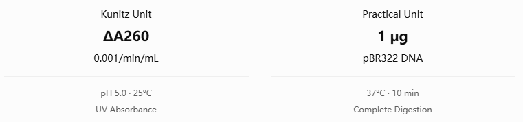
The classical Kunitz unit is defined by the absorbance increase at 260 nm as DNA is hydrolyzed: one Kunitz unit produces a ΔA260 of 0.001 per minute per mL at pH 5.0 and 25 °C in a defined sodium acetate–magnesium sulfate buffer [7][8]. The measurement relies on the hypochromic effect — intact double-stranded DNA absorbs less at 260 nm than its free bases, so degradation raises absorbance.
Most suppliers instead define the unit by digestion of a fixed amount of DNA under application-relevant conditions. In the HYJ525 manual, one unit is the amount that completely degrades 1 µg of pBR322 DNA in 10 minutes at 37 °C in DNase I reaction buffer — and the manual states the conversion explicitly: one DNase I unit equals 0.3 Kunitz units [6].
That single sentence is unusually useful, because it lets a specification written in Kunitz units be converted into the unit your assay actually measures. Without it, comparing a supplier’s “50 KU” against an in-house activity result is guesswork.
Three rules prevent most unit confusion: state the unit definition in the specification, not just the number; state the substrate and temperature, since Kunitz conditions (pH 5.0, 25 °C, genomic DNA) differ from process conditions (neutral pH, 37 °C, probe substrate); and never convert between vendors’ units without a documented conversion, because cross-vendor equivalence is an assumption, not a fact.
Running the Assay: Step by Step
Fix your own volumes, dilutions and acceptance criteria in a written procedure before routine use.
Step 1: Equilibrate the kit and prepare the probe
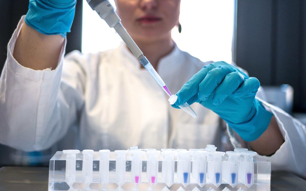
Bring the kit to 18–25 °C, mix the components and spin briefly. Reconstitute the DNA probe with 40 µL of TE buffer, aliquot to avoid repeated freeze–thaw, then dilute 50-fold with TE buffer to make the working solution. Protect it from light [6].
Step 2: Set the gain before the first run
This is the step most often skipped and most often responsible for poor results. Read a blank well and a well containing 1 µL of DNase I standard (2 U/μL) plus 79 µL of DNase & RNase-free water, incubate 30 minutes at 37 °C in the dark, then fix the gain — auto-scale (recorded as G1) or a medium setting adjusted up or down (recorded as G2) so the high signal sits inside the instrument’s linear range [6].
Step 3: Prepare controls and samples
The kit supplies a DNase I standard at 2 U/μL. A three-step dilution gives a positive control at 2×10⁻⁵ U/μL, with DNase & RNase-free water as the negative control [6]. Sample volume is 80 µL; dilute smaller samples to 80 µL with DNase & RNase-free water.
Step 4: Dispense, read RFU0, incubate, read RFU30
Add 10 µL of probe working solution and 10 µL of 10× reaction solution per well, then 80 µL of sample or control, in duplicate. Read immediately to record RFU0, incubate in the dark at 37 °C for 30 minutes, then read RFU30. Kinetic mode — reading every 1–1.5 minutes — is more informative than an endpoint when activity is low [6].
Step 5: Interpret the ratio, then quantify if needed
For screening, RFU30 ≥ 2 × RFU0 indicates DNase contamination [6]. For a concentration value, continue to the standard curve below.
Reading and Quantifying the Result
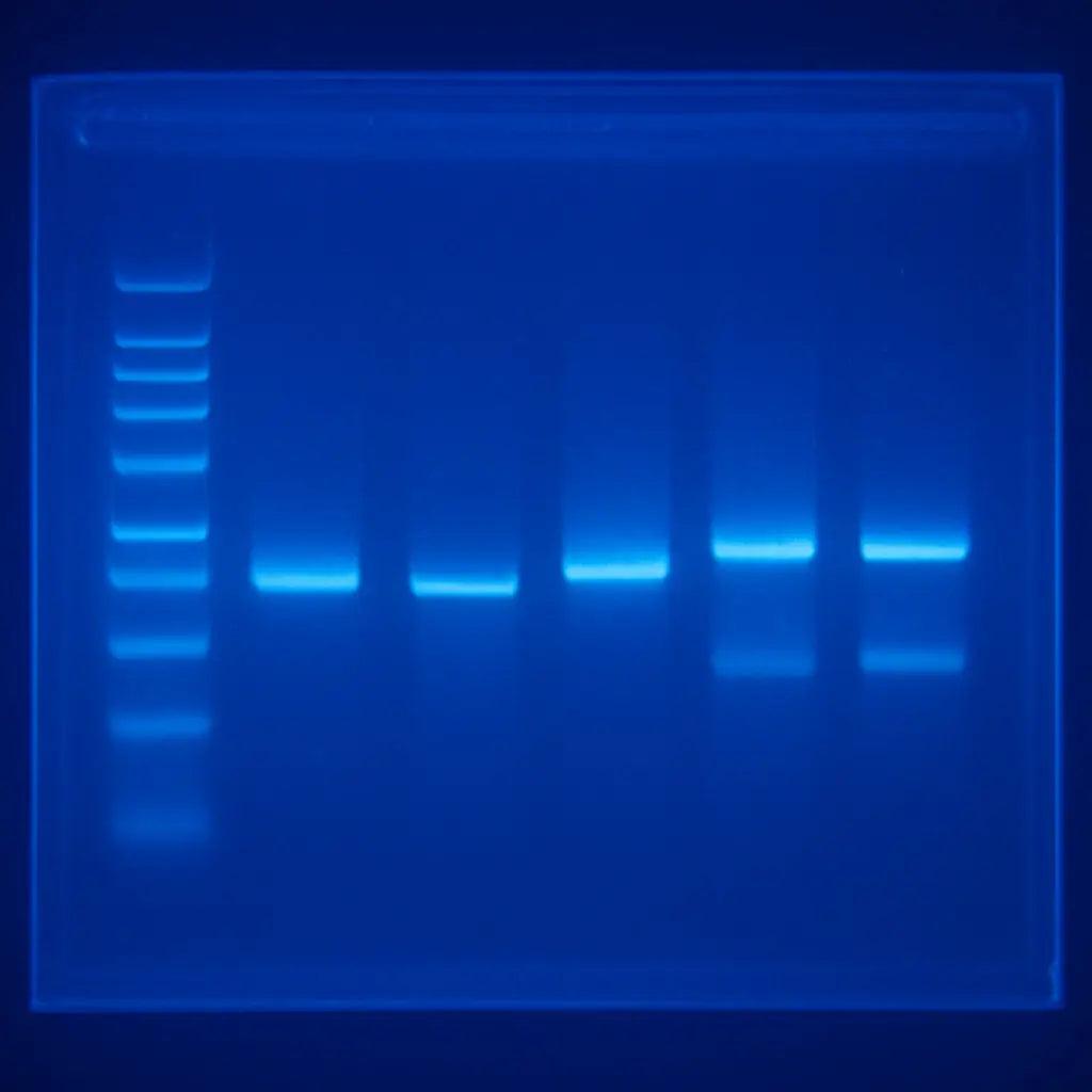
The 2× ratio is a practical cut-off, not a formal limit of detection, and two manual caveats matter. Heavily contaminated or interfering samples can produce false negatives — if RFU0 of the sample already exceeds RFU0 of the positive control while RFU30 stays below 2 × RFU0, pre-dilute and re-test rather than reporting negative. And matrix effects are real: dark, viscous or surfactant-containing samples interfere with fluorescence, while high ionic strength, pH below 4 or above 9, and protein denaturants inhibit the enzyme, so the result reflects the overall activity of the diluted sample rather than intrinsic enzyme activity [6].
For quantification, the manual provides ten standard levels from 1.25×10⁻⁶ to 1×10⁻⁵ U/μL plus a zero-concentration sample. Plot ∆RFU (RFU30 − RFU0) against concentration, fit a line, and require r ≥ 0.99 before use; a negative ∆RFU from signal fluctuation is treated as zero [6].
| Parameter | Stated value (HYJ525 manual) |
| Detection limit | 1.25×10⁻⁶ U/μL DNase I |
| Intra-batch precision | CV ≤ 10% |
| Inter-batch precision | CV ≤ 15% |
| Quantification range | 1.25×10⁻⁶ to 1×10⁻⁵ U/μL, r ≥ 0.99 |
| Format | 48 T / 192 T, black 96-well plate, Ex/Em 485/525 nm |
These describe kit performance, not a method validated for your sample matrix.
Validating the Assay Under ICH Q2(R2)
ICH Q2(R2), adopted in 2023, frames validation around the intended use of the procedure: a limit test needs specificity and a detection limit, while a quantitative impurity test needs specificity, accuracy, precision, range, linearity and a quantitation limit [9][10]. A DNase activity assay used for release or in-process control is usually the second type.
| Validation element | How to demonstrate it |
| Specificity / selectivity | Spike DNase into your real matrix and confirm the response is enzymatic; include a heat-inactivated or EDTA-chelated control to show the signal disappears when activity is blocked |
| Accuracy | Spike recovery at three levels across the range, three replicates each; report mean recovery with confidence interval |
| Repeatability | ≥ 6 determinations at one level in a single run; the kit’s CV ≤ 10% is a starting point, your matrix may be worse |
| Intermediate precision | Different day, analyst and plate lot; the kit’s CV ≤ 15% indicates the expected order of magnitude |
| Quantitation limit and range | Establish the lowest level with acceptable accuracy and precision, then show linearity across the working range; the 1.25×10⁻⁶ U/μL detection limit is a floor, not the LOQ |
| Robustness and false negatives | Vary incubation time, temperature and gain deliberately; spike negative samples post-hoc to prove the matrix does not mask activity |
Two operational points are easy to overlook. Nuclease-free consumables are part of the method — tips, tubes and plates must be certified, and the kit includes a decontamination reagent (DNase RNase away) applied to benches, gloves and surfaces for 5 minutes before wiping [6]. And because the assay measures activity, an inactivated enzyme reads as negative: a negative result does not prove the protein is absent. Where the control strategy needs both, pair the activity assay with an immunoassay.
Troubleshooting DNase Activity Assays
| Problem | Possible cause | What to try |
| No signal in the positive control | Probe not reconstituted or over-diluted; standard thawed repeatedly; gain too low | Prepare fresh probe working solution; use a fresh standard aliquot; repeat gain setting |
| Negative wells also rise | Nuclease contamination of water, tips, plate or bench | Use certified nuclease-free consumables; decontaminate surfaces; repeat with fresh water |
| Negative sample reads positive | Genuine contamination, or dye/surfactant interference | Pre-dilute and re-test; confirm with a spike-recovery control |
| Positive sample reads negative | Matrix inhibition (salt, pH < 4 or > 9, denaturants) or signal saturation | Pre-dilute with DNase & RNase-free water; re-check gain; re-test a spiked aliquot |
| High replicate variability | Small volumes, bubbles, edge effects, uneven temperature | Low-retention tips; brief spin; keep critical samples off outer wells; pre-warm the reader |
| Standard curve r below 0.99 | Dilution error, expired probe, or reading outside the linear range | Rebuild the dilution series; check probe storage; narrow the calibration range |
How to Choose a DNase Activity Assay Kit
- Detection limit against your control limit. The assay should sit well below the acceptance criterion to leave room for dilution and matrix effects. The HYJ525 detection limit is 1.25×10⁻⁶ U/μL.
- Whether quantification is supported. Screening needs a cut-off; specifications need a calibration curve with a standard of stated unit definition.
- Stated precision. Intra- and inter-batch CV values let you budget method variability before validation.
- Matrix guidance. Instructions on inhibitors, interfering substances and pre-dilution are the practical difference between a research kit and a QC tool.
- Contamination control built in. Nuclease-free water and a decontamination reagent reduce handling false positives.
- Format, throughput and documentation. Confirm the filter set (Ex/Em 485/525 nm), plate type and throughput, then ask for the manual, lot-specific QC data and intended-use labeling. Bulk and OEM/ODM terms matter if the kit supports your own product line.
Hyasen DNase Activity Assay and Process Impurity Products
Hyasen Biotech supplies process-related impurity detection kits and nuclease raw materials, with bulk supply and OEM/ODM support for diagnostic and biopharma manufacturers.
- DNase Assay Kit (Fluorescence) (HYJ525) — fluorescence probe format for DNase contamination screening and quantification. Detection limit 1.25×10⁻⁶ U/μL DNase I, intra-batch CV ≤ 10%, inter-batch CV ≤ 15%; 48 T and 192 T, Ex/Em 485/525 nm, 30-minute endpoint or kinetic read.
- RNase Assay Kit (Fluorescence) (HYJ526) — the same probe principle applied to RNase, using an RNA probe read at Ex/Em 535/575 nm; 48 T and 192 T. The paired screen for mRNA workflows, where RNase contamination is the mirror-image risk.
- DNase I ELISA Kit (HYJ527) — immunoassay format for DNase I protein, complementary to the activity assay when the control strategy requires both activity and antigen data. Refer to the product page for specifications.
- Additional process-related impurity assays for mRNA manufacturing: RIG-I dsRNA Detection Kit (HYJ522), T7 RNA Polymerase ELISA Kit (HYJ523) and (HYJ524).
- The enzymes these assays measure, available as bulk raw materials: Recombinant DNase I (RNase-free), Lyophilized Powder (HYC127) and DNase I (HYJ234).
Please refer to each product page and manual for specifications, and contact us about samples, bulk supply and OEM/ODM customization.
Frequently Asked Questions
What is a DNase activity assay?
A DNase activity assay measures how quickly a DNase sample degrades a defined DNA substrate. The substrate is incubated with the sample under fixed buffer and temperature conditions, and the rate of degradation is converted into activity units.
How does a fluorescence DNase activity assay work?
A quenched DNA probe carries a fluorophore and a quencher. When DNase cleaves the probe, the fluorophore is released and fluorescence increases, so rising signal tracks enzyme activity. The reaction is read at Ex/Em 485/525 nm in a microplate reader.
What does RFU30 ≥ 2 × RFU0 mean?
It is the qualitative cut-off in the HYJ525 manual: if fluorescence at 30 minutes is at least twice the time-zero reading, the sample is considered contaminated with DNase. Samples that are heavily contaminated or contain interfering substances should be pre-diluted and re-tested.
What is the detection limit of a fluorescence DNase assay?
For HYJ525, the stated detection limit is 1.25×10⁻⁶ U/μL DNase I, with a quantification range of 1.25×10⁻⁶ to 1×10⁻⁵ U/μL and a required correlation coefficient of at least 0.99. Other kits and other matrices will differ.
How do DNase units convert to Kunitz units?
The HYJ525 manual states that one DNase I unit — the amount that completely degrades 1 µg of pBR322 DNA in 10 minutes at 37 °C — equals 0.3 Kunitz units. Conversion factors are product-specific; do not apply them to another supplier’s enzyme.
Why is DNase testing required for biopharmaceutical products?
Nuclease is a process-related impurity. ICH Q6B requires such impurities to be controlled, and pharmacopoeial general chapters for vaccines, recombinant monoclonal antibodies and gene therapy products require nuclease residues to be tested. For mRNA, WHO guidance describes DNase digestion of the DNA template and requires removal of production enzymes to be demonstrated.
Does a negative DNase activity result prove the enzyme is absent?
No. An activity assay detects catalytic function, so an inactivated or inhibited enzyme reads as negative. Where the control strategy requires both, pair the activity assay with an immunoassay for the DNase protein.
Can a fluorescence DNase assay be used in a GMP laboratory?
The kit provides performance characteristics — detection limit, precision, calibration range — that support method validation, but each laboratory must validate the procedure for its own matrix and intended use under ICH Q2(R2). Confirm the intended-use labeling of the kit before including it in a GMP control strategy.
Conclusion
A DNase activity assay converts an invisible contamination risk into a number that can be specified, validated and trended. For biopharma manufacturers the assay has two jobs — proving an intentional nuclease step worked, and proving the nuclease is gone — and both rest on the same fundamentals: a defined substrate, a known unit definition, controlled conditions, and a result read against a validated criterion. Choose a format sensitive enough for your control limit, validate it in your own matrix, and treat consumables and decontamination as part of the method rather than housekeeping.
Evaluating a DNase activity assay for process development or QC release? Contact Hyasen to discuss samples, technical specifications, bulk supply, OEM/ODM and customized solutions.
References
- ICH Q6B, Specifications: Test Procedures and Acceptance Criteria for Biotechnological/Biological Products — process-related impurities, including enzymes used in manufacture, must be controlled.
- Chinese Pharmacopoeia 2020, general chapters on human vaccines, human recombinant monoclonal antibody products and human gene therapy products — nuclease listed among process-related impurities requiring testing in an appropriate intermediate.
- World Health Organization, Evaluation of the quality, safety and efficacy of messenger RNA vaccines for the prevention of infectious diseases: regulatory considerations (2022) — DNase digestion of the DNA template after IVT; removal of production enzymes to be demonstrated.
- WHO Technical Report Series 978 — residual cell-substrate DNA limit of 10 ng per parenteral dose for products from continuous cell lines.
- Kanaya S. et al., Highly sensitive nuclease assays based on chemically modified DNA or RNA, Sensors 2014, 14(7), 12437–12450 — fluorogenic probe principles and comparative detection limits.
- Hyasen Biotech, DNase assay Kit (Fluorescence) (HYJ525) manual — principle, components, storage, workflow, interpretation, performance characteristics and unit conversion.
- Kunitz M., Journal of General Physiology 1950, 33, 349–362 and 363–377 — original DNase I assay and unit definition.
- MilliporeSigma, Enzymatic Assay of Deoxyribonuclease I (EC 3.1.21.1), technical protocol — Kunitz assay conditions (pH 5.0, 25 °C, 83 mM sodium acetate, 4.2 mM MgSO₄).
- ICH Q2(R2), Validation of Analytical Procedures (2023) — validation characteristics by intended use.
- ICH Q14, Analytical Procedure Development (2023) — analytical target profile and lifecycle approach.